Breast Cancer Surgery
What is Breast Surgery?
The purpose of breast cancer surgery is to remove the cancer while balancing the effects on appearance, sensation and function. The treatment plan for the cancer should be personalised to meet the demands and expectations of each patient. Your breast cancer specialist should discuss these choices with you and help you agree on the best treatment option for you.

Breast cancer is the most common cancer in women. Early detection through screening is important. If a cancer is found in its early stages, it can increase the chances of successful treatment.
Clinical Breast Examination
Have a doctor to examine your breasts once every year if you are 40 years and above. This includes a visual examination and a manual check of the entire breast and underarm area for changes. Changes in the breast may not be due to cancer and diagnostic tests may be performed to assess these changes.
Mammogram Screening
Mammography is a low-powered X-ray technique that gives an image of the internal structure of the breast. Usual screening mammograms involve taking X-ray images with the breast compressed between two plates with two views taken — cranial caudal or horizontal and mediolateral oblique or diagonal.

Additional angles and magnified views may be taken if there are areas of concern. It can detect the presence and position of abnormalities and help in the diagnosis of breast problems including cancer.
The risk of developing breast cancer increases with age. Women with risk factors such as a family history of breast cancer should discuss with their doctors when to go for and the interval of regular screening.
There are other tests such as breast ultrasound, tomosynthesis and MRI, available for assessment of the breasts but these are not used for regular screening in well women and are used for further evaluation after initial screening mammogram but may be considered for women with high risk of breast cancer.
After diagnostic tests reveal that your breast lumps are cancerous, the next step of treatment is surgery.
Minimally Invasive Breast Biopsies
A lump in your breast may not necessarily mean cancer. To determine, whether a lump is benign or malignant, biopsies are sometimes performed to enable the doctor to study the tissue and determine if it is cancerous.
Fine Needle Aspiration (FNA)
A syringe with a very fine needle is used to withdraw fluid or cells from a breast lump. This is a simple procedure and can be uncomfortable but is usually tolerable enough for it to be done in the clinic.
If the lump is just a cyst, withdrawing fluid in this manner will usually make the cyst disappear.
However, if the lump is solid, your doctor may use this procedure to withdraw some cells from it. The cells will then be sent to a laboratory for examination.
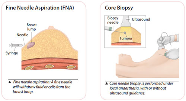
Core Needle Biopsy
This is a minimally invasive method that obtains a few tiny strips of tissue from an area of abnormality with a wide bore needle. Local anaesthetic is injected to numb the breast area, followed by a small incision in the skin to allow easy insertion of the needle.
If the abnormality is non- palpable (not detectable by clinical examination) and visible on the ultrasound, ultrasound guidance is used to obtain the tissue. Usually 2 to 6 cores of tissue will be obtained for examination.
A nurse will apply compression to the breast to stop any bleeding. The wound is closed by a steristrip and the dressing applied. Strenuous activity is to be avoided for 2 days after the biopsy.
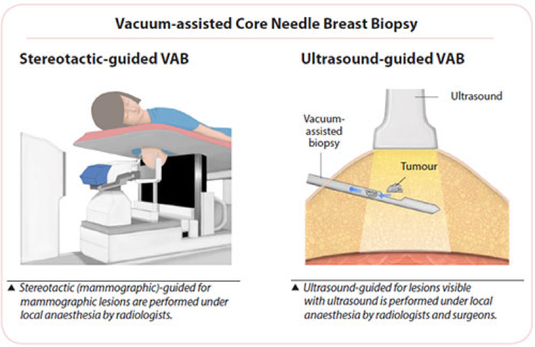
Vacuum-assisted Core Needle Breast Biopsy
Vacuum-assisted biopsy (VAB) devices use a larger bore needle with a vacuum component to obtain tissue samples from non-palpable lesions.
Like the usual core biopsy, this minimally invasive procedure is also performed under local anaesthesia, which is injected to numb the breast area, followed by a small incision in the skin to allow easy insertion of the needle. It is used for lesions seen by mammography (stereotactic-guided biopsy), ultrasound or MRI.
The surgeon or radiologist places the probe into the suspicious area of the breast accurately. A vacuum then draws the tissue into the probe, a cutting device removes the tissue sample and then carries it through the probe into a collection area. More tissue is usually obtained using the VAB than the usual core needle biopsy and the number of strips removed is dependent on the area that needs to be examined.
A small titanium clip (microclip) may be placed at the biopsy site as a location marker for future treatment. This clip is very small (2 mm), is harmless, and will not cause any problems when left inside the breast. An X-ray is taken post-biopsy to ensure proper clip placement. New biodegradable markers are also available now.
A nurse will apply compression to the breast to stop any bleeding, the wound is closed by a steristrip and the dressing applied. Strenuous activity is to be avoided for 2 days after the biopsy.
This procedure is minimally invasive as compared to an open surgical biopsy. It is performed as a day surgery procedure. lt has the ability to sample tiny abnormalities called microcalcifications, making early diagnosis of breast cancer possible.
Types of Surgery
There are generally two types of surgery — lumpectomy (breast conservation surgery) and mastectomy.
Lumpectomy (Breast Conservation Surgery)
Lumpectomy is a surgical procedure to remove only a portion of the breast containing cancerous tissues while preserving the rest of the healthy breasts.
Radiation therapy is usually provided after a lumpectomy to remove any remaining cancer cells left in the breast to minimise the risk of cancer returning.

WLE-with-Axillary-Dissection
Level-2-Axxilary-Clearance
Mastectomy
The entire breast tissue is surgically removed from the breasts.
The different types of mastectomy include:
Total Mastectomy
Involves removing the entire breast, including the nipple, the circular area around the nipple (areola) and most of the overlying skin. Usually performed when the cancer has not spread beyond the breast.

Advanced Breast Cancer
Mastectomy with Axillary Clearance
Radical Mastectomy
Similar to total mastectomy. However, the chest muscles under the breast and lymph nodes are also removed. This surgery is advised when the cancer has spread to the chest muscles.
Skin-Sparing Mastectomy
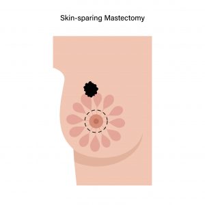
The entire breast tissues, nipple and areola are removed, but most of the skin over the breast remains intact. It is usually performed if a breast reconstruction surgery is planned immediately after mastectomy.
However, it may not be recommended if the tumour is too large or near the skin surface.
Nipple-Sparing Mastectomy
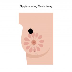
Similar to skin-sparing mastectomy. However, the breast tissues are only removed up to the nipple and areola. The skin of the nipple and areola are preserved, and the tissues under and around them are removed.
If the tissues removed turn out to be cancerous, the skin of the nipple and areola must also be removed.
A breast reconstruction surgery is done after this surgery.